US Görüntüleme / Temporomandibular Eklem anatomisi, patolojileri, görüntüleme ve tedavileri
Uygulamalı Kurs
Kurs tarihi ve yeri: 14-17 Nisan 2022
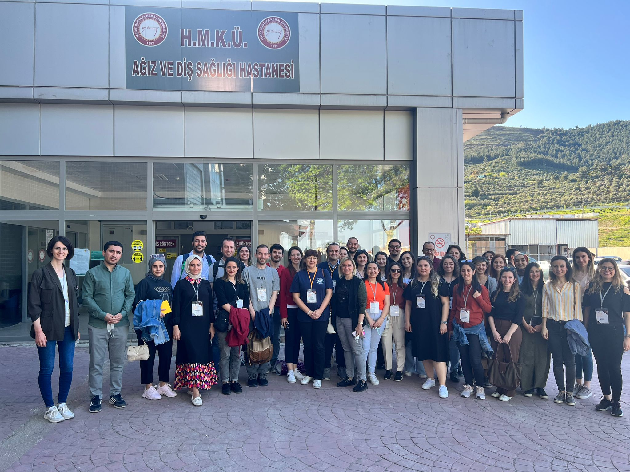
Hatay Mustafa Kemal Üniversitesi, Atatürk Konferans Salonu
HMKÜ, Diş Hekimliği Fakültesi, Ağız, Diş ve Çene Cerrahisi Kliniği
Prof. Dr. Ingrid Rozylo-Kalinowska
Dr. Kaan Orhan
Doç. Dr. Hakan Eren
Doç. Dr. Gözde Serindere
Özellikle farklı patolojiler ve sistemik durumlarla birlikte maksillofasiyal bölgenin anatomisi ve patolojisine aşina olmak, klinisyenlerin özellikle ağrının eşlik ettiği gizli hastalıkları ve maksillofasiyal patoloji vakalarını keşfetmesine yardımcı olacaktır.
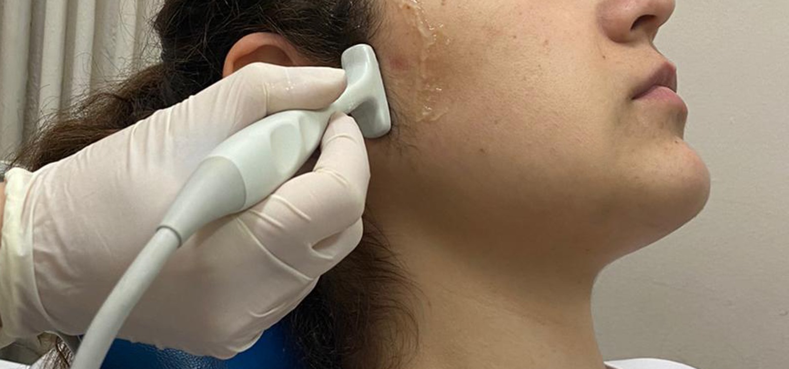
Ultrasonografi (USG), klinisyenler tarafından yeterince kullanılmayan bir araştırma aracıdır. Hastalar için yerinde teşhis sağlayabilir ve ucuzdur. Maksillofasiyal USG, özellikle intraoral uygulamalar nispeten yenidir ve maksillofasiyal radyologlar için birçok potansiyel kullanım sağlar. Operasyon sırasında sadece tiroid nodülleri dahil boyundaki şişliklere bakmak için değil, aynı zamanda dil, oral kavitenin yumuşak dokusu gibi yapılara bakmak için de çok faydalı bir araçtır.
Bu kurs Orofasiyal Ağrı ve Temporomandibular eklem (TME) Bozukluklarının tanı ve tedavisi ile ilgili bilgileri sunmak için tasarlanmıştır. Kurs, bir dizi ders ve vaka sunumlarından oluşacaktır. Bu kursta verilen bilgiler, katılımcıların, diş hekiminin karmaşık orofasiyal ağrı problemlerini yönetmedeki rolünü anlamalarını sağlayacaktır. Klinisyenler, bu ağrı bozukluklarının tedavisinde önemli bir rol oynadığından, temporomandibular hastalıklar konusu üzerinde durulacaktır. Maksillofasiyal patolojiler için tedavi planlaması, mümkün olduğunca fazla bilgi toplamayı içerir. Bu nedenle, dersin odak noktası ile birlikte, USG ve manyetik rezonans görüntüleme (MRI) üzerinde özellikle durularak, TME'nin çeşitli modalitelerle görüntülenmesi de tartışılacaktır.
Öğrenme hedefleri:
1. USG'yi etkili bir şekilde nasıl kullanacağını bilmek
2. USG'de görülen özellikleri anlamak
3. Baş ve boyun USG uygulamasını öğrenmek
4. Tükürük bezi hastalığını anlamak
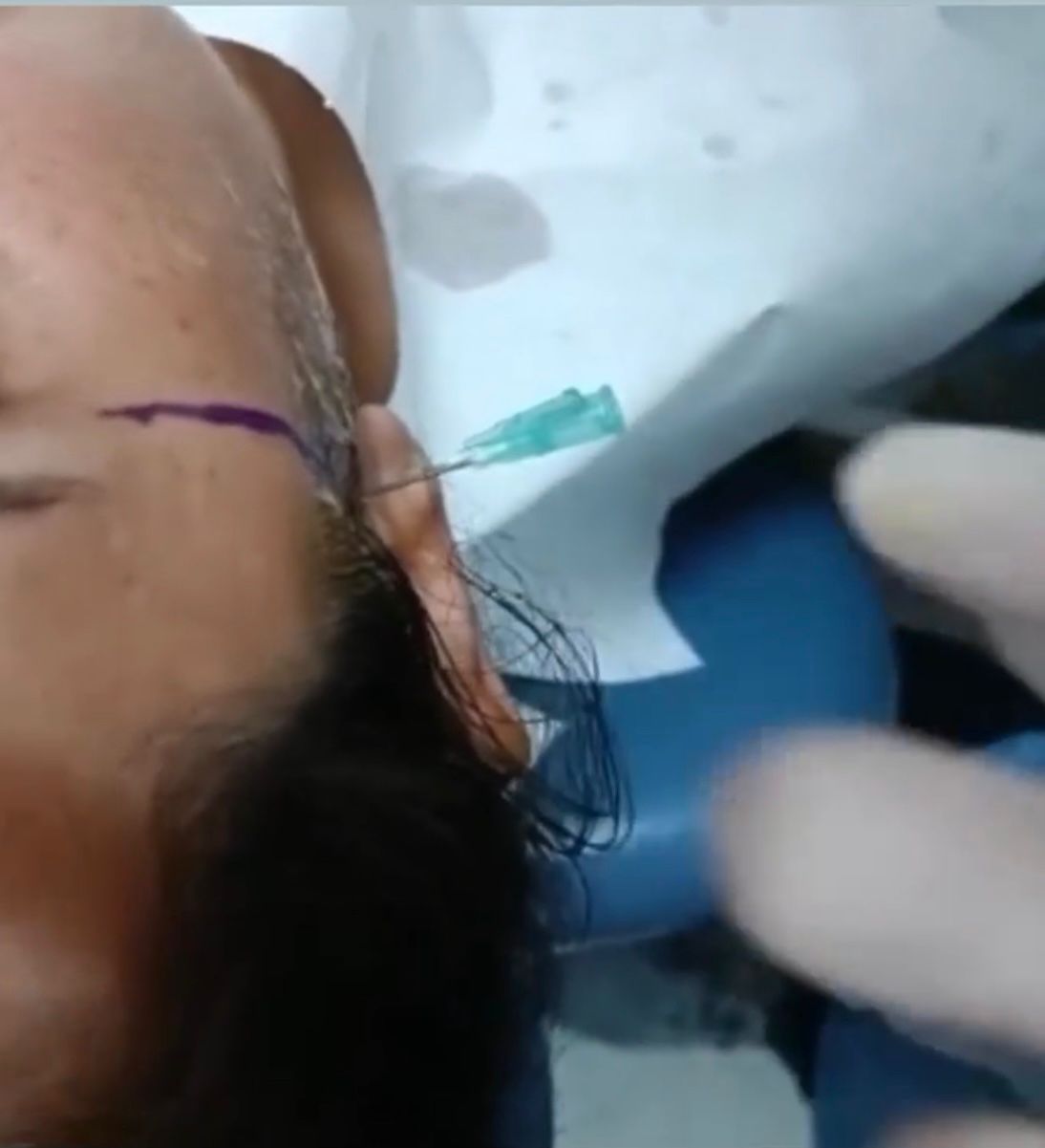
5. USG rehberliğinde ince iğne ve kor biyopsisinin nasıl yapıldığını öğrenmek
Sunumdan sonra katılımcılar şunları yapabileceklerdir:
• Uygun görüntüleme yöntemine olan ihtiyacı değerlendirebilecek, hastaları için uygulanacak görüntüleme yöntemini açıklayabileceklerdir.
• USG ile yumuşak dokuların ve TME'nin görüntülenmesinde ne gerekli, ne değil?
Program
14 Nisan 2022- Perşembe
12:00-13:00 Kayıt
13:00-13:10 Giriş
13:10-13:45 USG’nin temel esasları-- Prof. Dr. Ingrid Rozylo- Kalinowska
13:45-14:15 Baş ve Boyun Anatomisi ve fizyolojisi /TME-- Prof. Dr. Ingrid Rozylo-Kalinowska
14:15-14:45 ABD Baş ve boyun Anatomisi -- Doç. Dr. Hakan Eren
14:45-15:15 Çay/kahve arası
15:15-16:30 Maksillofasiyal Görüntülemede USG Kullanımı-- Doç.Dr. Hakan Eren
16.30-17:00 Çay/kahve arası
17:00-18:00 TME Görüntülemeye Giriş-- Doç. Dr. Gözde Serindere
18:00 Birinci Günün Sonu
15 Nisan 2022 Cuma
09:00-10:00 TME değerlendirmede Konik Işınlı Bilgisayarlı Tomografi -- Prof. Dr. Ingrid Rozylo-Kalinowska
10:00-11:00 TME değerlendirmede MRI/USG-- Prof. Dr. Kaan Orhan
11:00-11:30 Çay/kahve arası
11:30-12:30 TME patolojilerinin semptomları-- Prof. Dr. Kaan Orhan
12:30-13:30 Öğle Yemeği
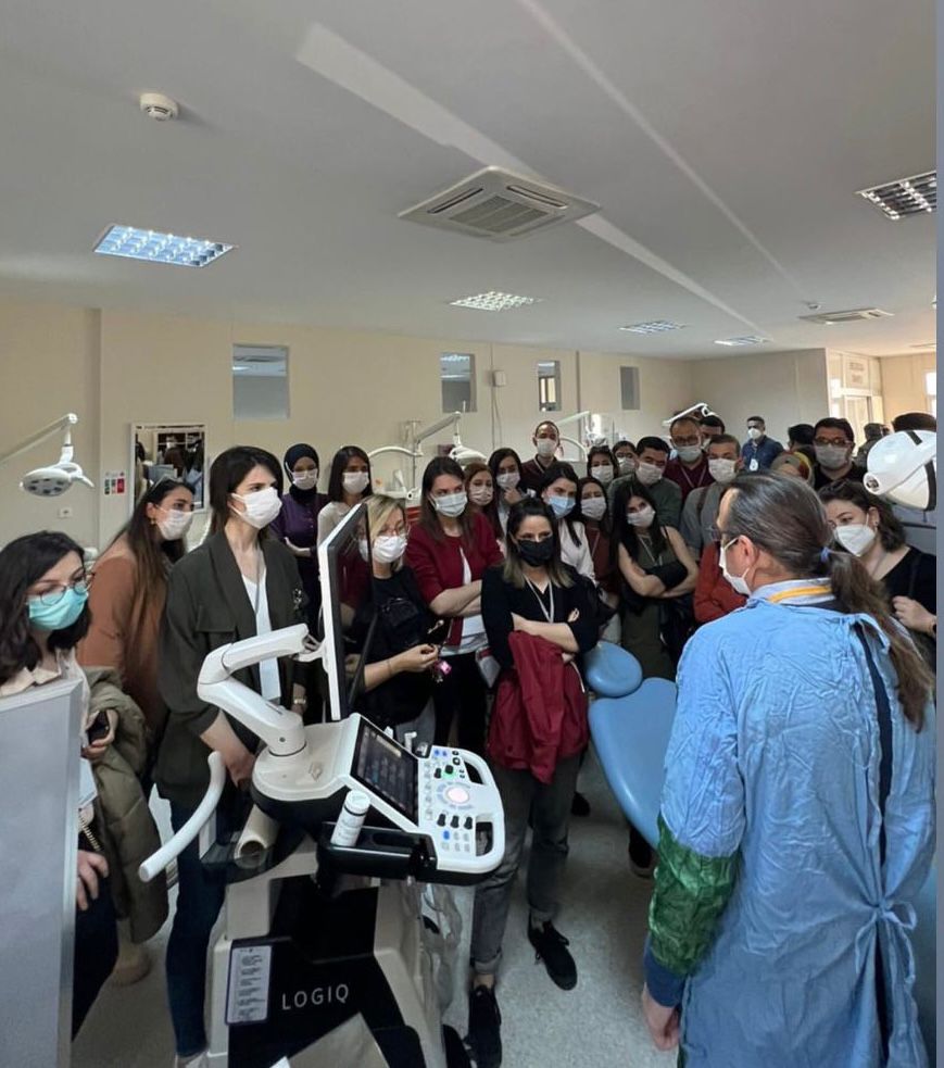
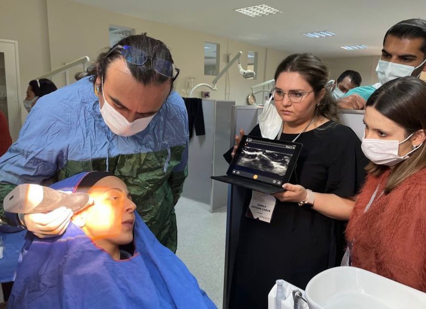
13:30-17:00 USG baş ve boyun/TME görüntüleme uygulamalı kurs-- Prof. Dr. Ingrid Rozylo-Kalinowska/ Prof. Dr. Kaan Orhan/ Doç. Dr. Hakan Eren
Sosyal Program
- Türkiye’nin en büyük mozaik müzesi olan Hatay Arkeoloji Müzesi gezisi
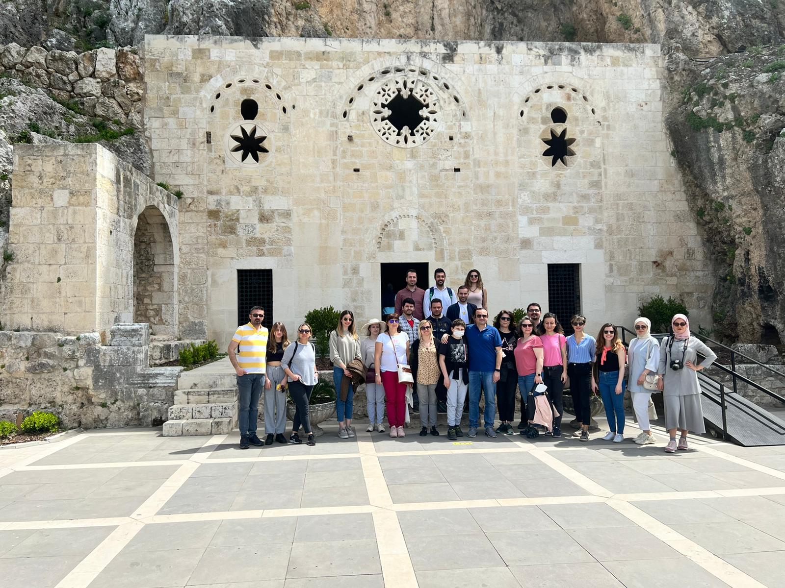
- “Dünyanın ilk mağara kilisesi” olarak kabul edilen St. Pierre Kilise ziyareti
- Öğle yemeği molası
- Hoşgörü kenti olarak bilinen Hatay’ın simgesi olarak gösterilen, cami, kilise, havra üçgeni arasında, önemli bir turistik yer olan uzun çarşı gezisi ve alışveriş
- Eski Antakya sokakları gezisi
Gala Yemeği
Gala yemeği, sosyal program sonrası Antakya Konak Restaurant’da gerçekleştirilecektir.
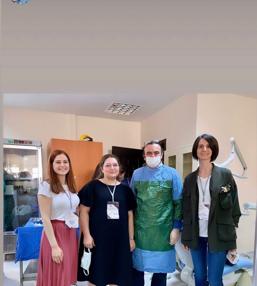
Etkinliğimizden Kareler
